Project Goal
To develop advanced imaging technologies that will be used to view and understand plant roots and below-ground interactions.
Project Summary
Project 2.3 is working to determine the best imaging technologies for high-throughput data acquisition, and which technologies have limitations. Emil Hallin and his team of researchers are using and developing two complementary tools in order to better understand and image a plant’s roots. The first tool is Phase Enhanced Imaging or Thermal Neutrons, and the second tool is Phase Enhanced Absorption and the laser-based Synchrotron Light. These are used to image the root microbiome or underground community and observe how the soil, roots, and microorganisms interact. This work will lead to the ability to conduct experiments and build a greater understanding of the unseen portion of a plant: its roots.

Project Results to Date
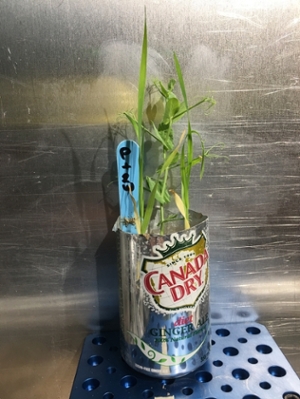
The Betatron Beamline in Montreal and Spectrometer N5 at the Canadian Neutron Beams Centre have been used to image plants both above and below ground, through 10 cm of soil. While other groups have done reconstructions, no one has successfully reconstructed root architectures for living plants in undisturbed soil media as thick as 8 cm. The researchers have also successfully digitally extracted roots from commercially used soil mixes, and are extending their work to allow imaging the rhizosphere and the organisms within it.
Colleagues in the College of Pharmacy and Nutrition are using an analytical technique called mass spectrometry to track nutrients within the roots of plants. The mass spectrometry facility led by Drs. Anas El-Aneed and Randy Purvus is in the process of developing methods to identify and characterize nutrients and nutrient bioproducts within the roots of plants.
Practical Applications
- Project 2.3 provides the opportunity to view images of intact root systems and the flow of water in the soil. This will lead to an increased ability to learn, observe, and understand the plant's underground community, as well as clearly observe interactions between roots and soil microorganisms. It will also enable further increases in plant productivity and adaptability to stressors. Understanding root architecture can lead to better methods of pulling nutrients into plants and ultimately make plants better and more efficient. This research may also lead to a decreased reliance on fertilizer, as scientists may have the ability to deliberately change root architecture to increase the efficiency of fertilizing methods.
- This research is changing how experiments are conducted by plant breeders and physiologists. The new digital tools being developed mean that plant roots do not have to be disturbed and soil does not have to be physically removed in order to view the roots. Soil can be digitally removed from images utilizing new technologies. This work is creating an exciting and dynamic opportunity to study living plants and how they interact with (and change) the rhizosphere in natural soils without disturbing any components of the rhizosphere.
- Implications of this research may also include how insecticides are used and affect the soil and the insects within it.
Collaborations
Project 2.3 utilizes technologies from the Advanced Laser Light Source, and Canadian Neutron Beam Centre. It also collaborates with the Global Institute for Water Security, the Controlled Environment Systems Research Facility, the University of Saskatchewan College of Pharmacy and Nutrition, and MS Facility.
Some of Project 2.3's collaborations with other P2IRC projects include:
- Project 3.4: Systems and Collaboration, a computing theme focused on collaboration and data sharing. Working with Project 3.4 has been particularly useful for Project 2.3 in its cross-country collaboration with other researchers.
- Project 3.2: Data Analysis for Rapid Plant Phenotyping, which focuses on creating tools for data analysis and collecting plant imagery. This project has developed software to aid Project 2.3’s image analysis capabilities.
Research Team
Loading...


